

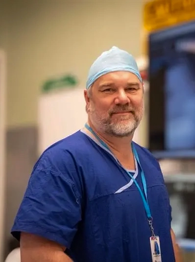
Authored by Dan Fick
Published on the Medical Forum online news channel, August 6, 2026
Orthopaedic care is moving from a focus on structure as cause of symptoms towards a more comprehensive model.
The orthopaedic care plan is a structured, patient-specific roadmap integrating advanced assessment, multidisciplinary treatment and, when required, surgical planning.
It brings together the expertise of the orthopaedic surgeon, general practitioner, physiotherapist, podiatrist, sports physician and other healthcare professionals to ensure every available treatment pathway is considered.
Advanced functional imaging, such as EOS imaging, plays an important role in helping create this plan by providing a more complete understanding of how a patient’s body functions under real-life conditions.
A whole-patient approach
Knee pain may be influenced by lower limb alignment, foot mechanics, muscle weakness, movement patterns or spinal and pelvic dysfunction. Similarly, hip symptoms may not be explained by the hip joint alone. An orthopaedic care plan considers:
- What is driving the patient’s symptoms?
- What factors can be modified without surgery?
- What treatments can optimise function and performance?
- If surgery is required, how can the patient be prepared to achieve the best possible outcome?
Advanced functional imaging
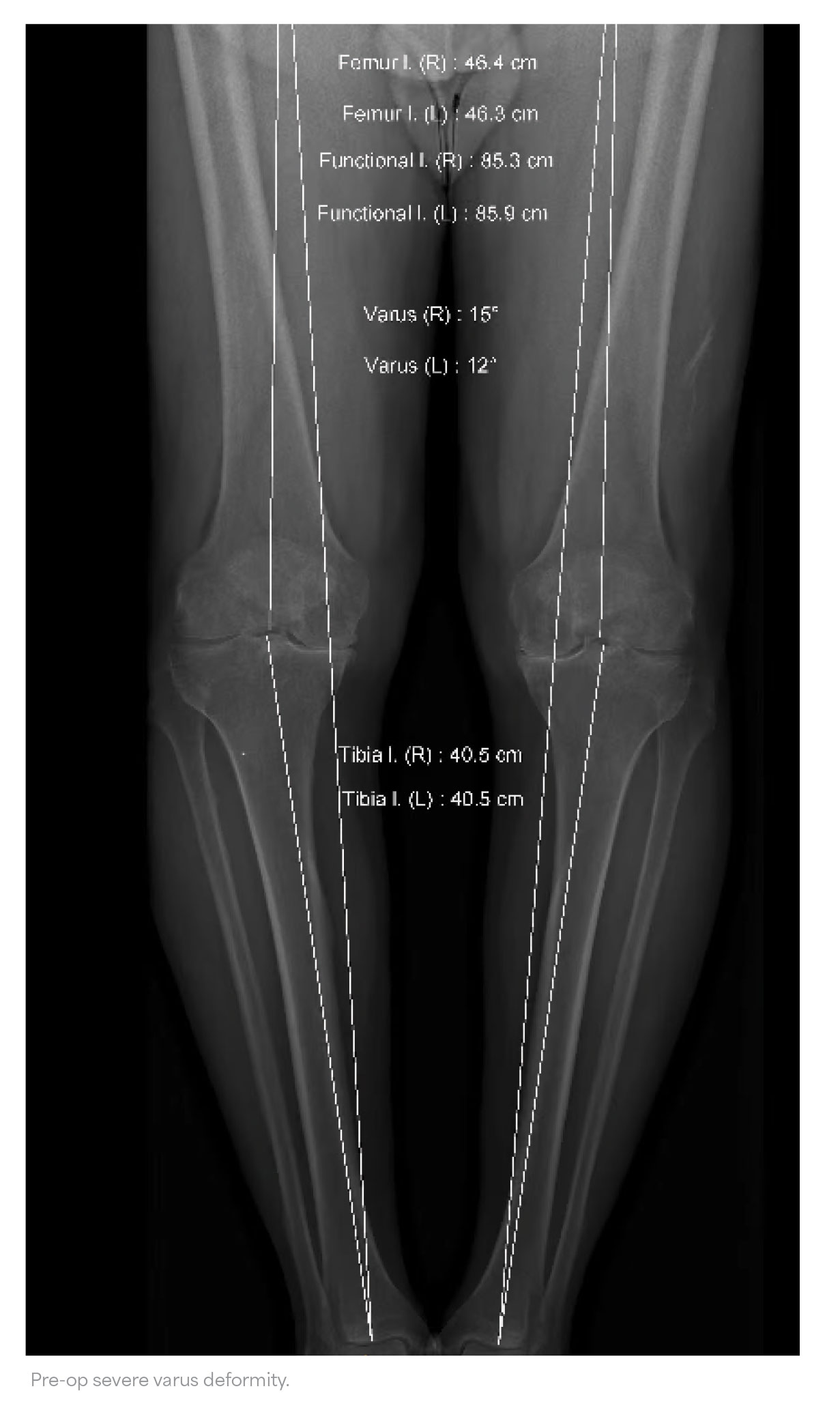
Traditional imaging remains an essential part of diagnosis. However, many investigations provide only a static view of anatomy. EOS functional imaging allows assessment in weight-bearing positions providing information about alignment and the interaction between different regions of the body.
This can help identify spinal and pelvic alignment patterns, lower limb mechanical alignment, functional changes between standing and sitting and biomechanical factors contributing to symptoms.
This is not only to determine whether a patient requires surgery, it is to help create a comprehensive treatment plan unique to the patient including optimising conservative management first.
Pre-surgery multidisciplinary pathway
Now we prioritise appropriate non-operative strategies and ensure they are targeted to the patient’s individual biomechanics. This usually involves close collaboration with:
- Physiotherapy: to address strength deficits, movement patterns, joint loading strategies, mobility and functional goals.
- Podiatry: to assess foot mechanics, gait abnormalities, orthotic requirements, and contribution of lower limb alignment to symptoms.
- Sports physicians: for patients requiring more complex management, including targeted and repeated injection therapies as well as diagnostic procedures to better understand pain sources.
For selected patients, emerging treatments such as radiofrequency ablation, radiotherapy and microembolisation procedures may form part of a broader strategy when appropriate.
Communication is key. The orthopaedic care plan is shared back with the GP, and other treating clinicians so that the entire healthcare team is working towards the same objectives.
The orthopaedic surgeon’s role is increasingly that of a care coordinator helping to integrate multiple specialties into a unified patient pathway.
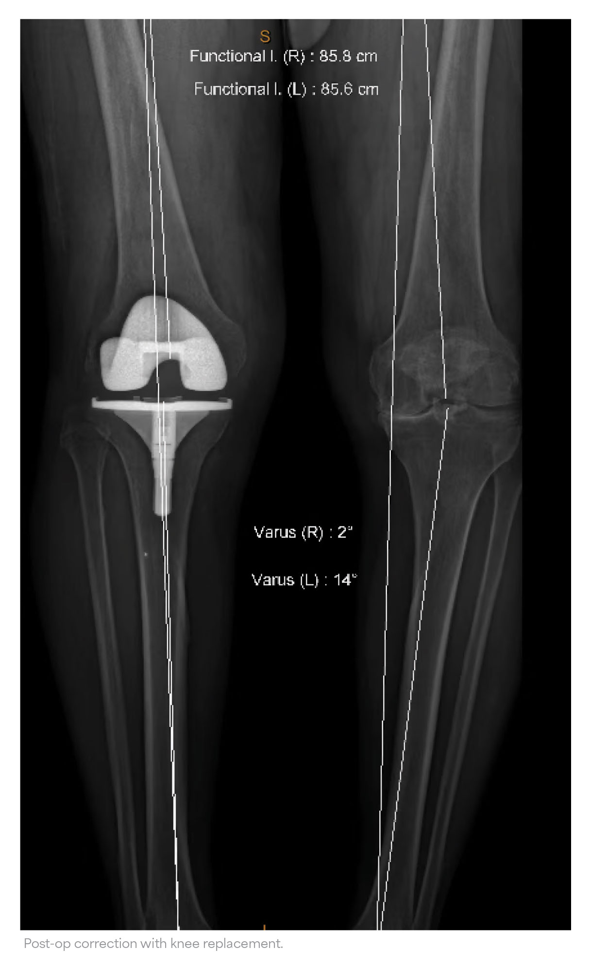
The same principles apply to patients progressing to a joint replacement; hip and knee arthroplasty outcomes depend not only on the implant, but on how that implant interacts with the patient’s anatomy and movement patterns. In joint replacements, the relationship between the spine, pelvis, hip and knee can influence implant stability.
Spinopelvic function and hip replacement
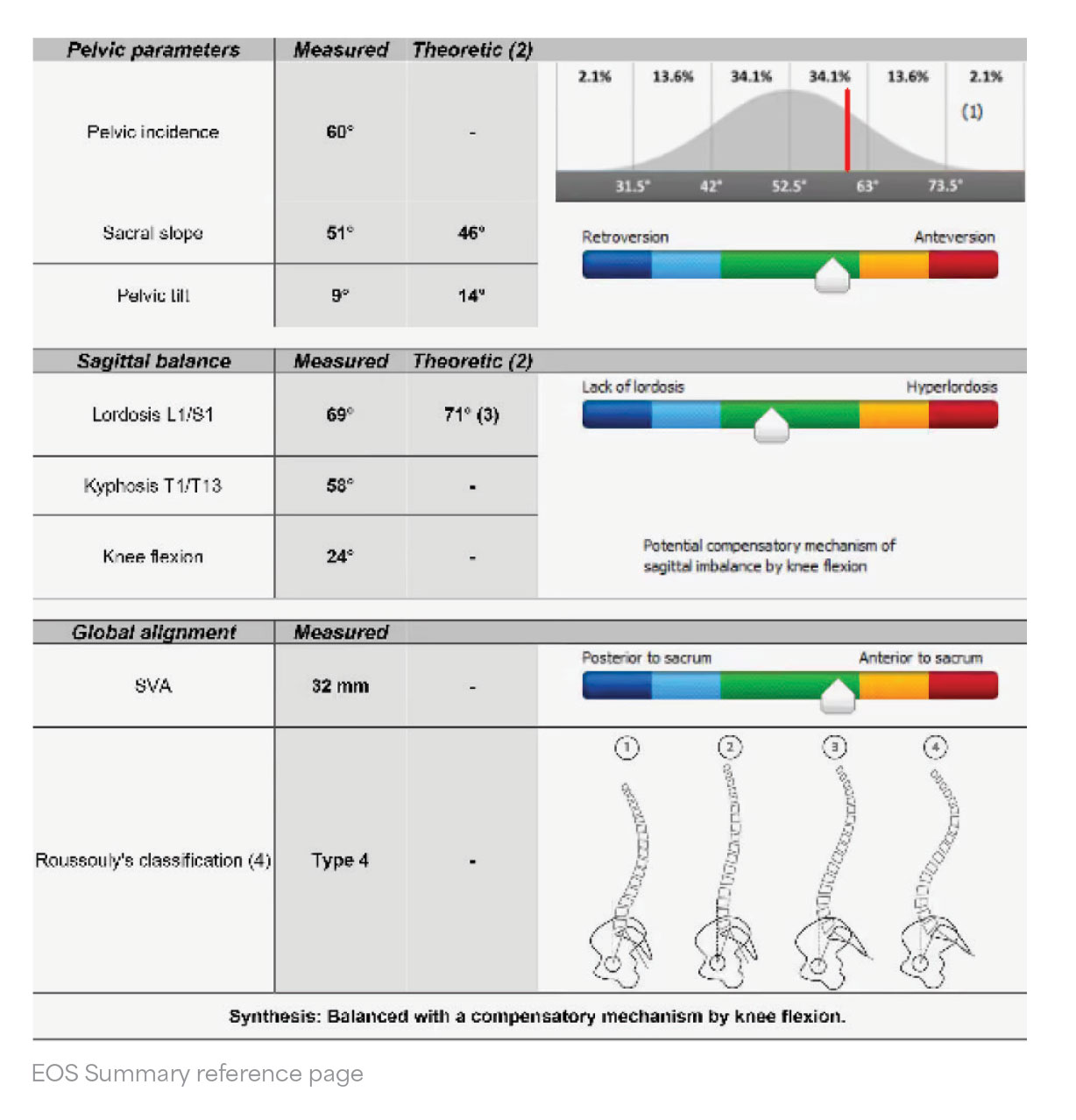
The spine and pelvis normally adapt during everyday movement. When a person moves from standing to sitting, the pelvis rotates to allow the hip joint to move efficiently. Spinopelvic dysfunction occurs when this relationship is altered, often due to spinal stiffness, degenerative spinal disease, previous fusion surgery, and abnormal pelvic positioning.
When the pelvis cannot adapt normally, the functional orientation of the hip socket may change between standing and sitting. This can increase the risk of impingement and instability following hip replacement.
EOS functional imaging helps surgeons understand these dynamic relationships and incorporate them into surgical planning. In patients with increased instability risk, dual mobility hip replacement technology provides another option.
A dual mobility hip replacement incorporates a mobile polyethylene liner that articulates within the metal acetabular component while also articulating with the femoral head. This design increases the effective head size and improves the stability characteristics of the implant.
They may be particularly beneficial in selected patients with factors such as spinopelvic dysfunction, spinal stiffness, previous spinal surgery and increased risk of dislocation.
The decision to use dual mobility components is part of a broader personalised surgical strategy informed by the patient’s individual biomechanics.
Change is now
For GPs, referral to an orthopaedic surgeon is increasingly not just a step towards surgery, it is an opportunity to develop a coordinated management plan involving the entire healthcare team.
Gone are the days of the surgeon dismissing patients with the wave of a hand and saying, “you’ll know when you’re ready – come back when you can’t stand it any longer”.
Our job is to better prepare patients for surgery and optimise their current pre-surgical state.
The goal remains a simple one: to maximise function, reduce pain and achieve the best possible outcome for every patient. Surgery is often not mandated to achieve this.
Go to Medical Forum for further conversations with the people shaping medicine in Western Australia.
Medical Forum is the leading independent monthly for Health Professionals in WA.
